

Histochemical scoring assessment (H-score)
Poosit Ruengwanichayakun
Department of Pathology, Floor 6, Sirindhorn Building, Naresuan University Hospital,
99 Moo 9 Phitsanulok-Nakhonsawan Road, Tha-pho, Muang, Phitsanulok 65000 Thailand.
Telephone: +66 (0) 89 439 2640 Fax: +66 (0) 55 965 331 Email: poosit.rue@hotmail.com
An evaluation of immunohistochemical staining is performed on a light microscope first using 10x objective (magnification of 100x) in order to scan and locate the histopathological appearances. Then the 40x objective (magnification of 400x) is subsequently applied for more detailed information on the staining(1,2).
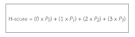
An interpretation of immunoreactivity is based on the histochemical scoring (H-score) assessment incorporating both the staining intensity (i) and a percentage of stained cells at each intensity level (Pi). The i values are indicated as 0 (no evidence of staining), 1 (weak staining), 2 (moderate staining), and 3 (strong staining). The Pi values vary from 0% to 100%. The final H-score is derived from the sum of i multiplied by Pi as the equation shown below. This score, therefore, is in the range of 0 to 300(3-5).
References
(1). Keller, E. & Goldman, R.D. 2006, "Light microscopy" in Basic Methods in Microscopy: Protocols and Concepts from Cells: A Laboratory Manual, eds. D.L. Spector & R.D. Goldman, Cold Spring Harbor Laboratory Press, China, pp. 1-42.
(2). Nicholson, R.I., Bouzubar, N., Walker, K.J., McClelland, R., Dixon, A.R., Robertson, J.F., Ellis, I.O. & Blamey, R.W. 1991, "Hormone sensitivity in breast cancer: influence of heterogeneity of oestrogen receptor expression and cell proliferation", European journal of cancer (Oxford, England : 1990), vol. 27, no. 7, pp. 908-913.
(3). McCarty, K.S.,Jr, Miller, L.S., Cox, E.B., Konrath, J. & McCarty KS, S. 1985, "Estrogen receptor analyses. Correlation of biochemical and immunohistochemical methods using monoclonal antireceptor antibodies", Archives of Pathology & Laboratory Medicine, vol. 109, no. 8, pp. 716-721.
(4). McCarty, K.S.,Jr, Szabo, E., Flowers, J.L., Cox, E.B., Leight, G.S., Miller, L., Konrath, J., Soper, J.T., Budwit, D.A. & Creasman, W.T. 1986, "Use of a monoclonal antiestrogen receptor antibody in the immunohistochemical evaluation of human tumors", Cancer research, vol. 46, no. 8 Suppl, pp. 4244s-4248s.
(5). Tadrous, P.J. 2007, "Breast" in Diagnostic Criteria Handbook in HISTOPATHOLOGY: A Surgical Pathology Vade Mecum John Wiley & Sons, England, pp. 258-266.